Noticing a small bump on your skin often leads to quick speculation: Is it a callus, a skin tag, or a viral growth? A wart is a non-cancerous skin growth caused by an infection in the top layer of the skin with the Human Papillomavirus (HPV). Because these growths can resemble several other common dermatological conditions, accurate visual identification is essential for choosing the right treatment and preventing unnecessary skin irritation.
Recognizing the distinct characteristics of different growth types, understanding where they typically appear, and identifying key markers—such as tiny black dots within the lesion—can help you determine what kind of skin lesion you are dealing with.
The Underlying Cause: What Makes a Growth a Wart?
Warts develop when HPV enters the epidermis through tiny micro-abrasions, cuts, or damaged skin barrier areas. The virus stimulates rapid keratinocyte proliferation in the outer skin layer, forming a raised, hyperkeratotic lesion.
Because HPV spreads through direct contact or contaminated moist surfaces (such as locker room floors, pool decks, or shared towels), these growths frequently appear on areas prone to minor trauma, such as fingers, knuckles, elbows, and feet.
Infection Pathway:
[Micro-abrasion in Skin] ➔ [HPV Penetrates Epidermis] ➔ [Keratinocyte Overgrowth] ➔ [Hyperkeratotic Lesion Forms]
Key Visual Identifiers of Common Warts
While different variants exist depending on the HPV strain and anatomical location, most viral growths share core visual features:
- Rough, Grainy Surface: Unlike smooth skin tags or soft moles, common viral lesions typically feature a rough, firm texture often compared to cauliflower.
- Distinct Border: The lesion usually has a clear, elevated edge separating it from normal surrounding skin.
- Interrupts Natural Skin Lines: Unlike calluses, where skin ridges (dermatoglyphics or fingerprints) run continuously across the bump, viral growths interrupt these natural skin lines.
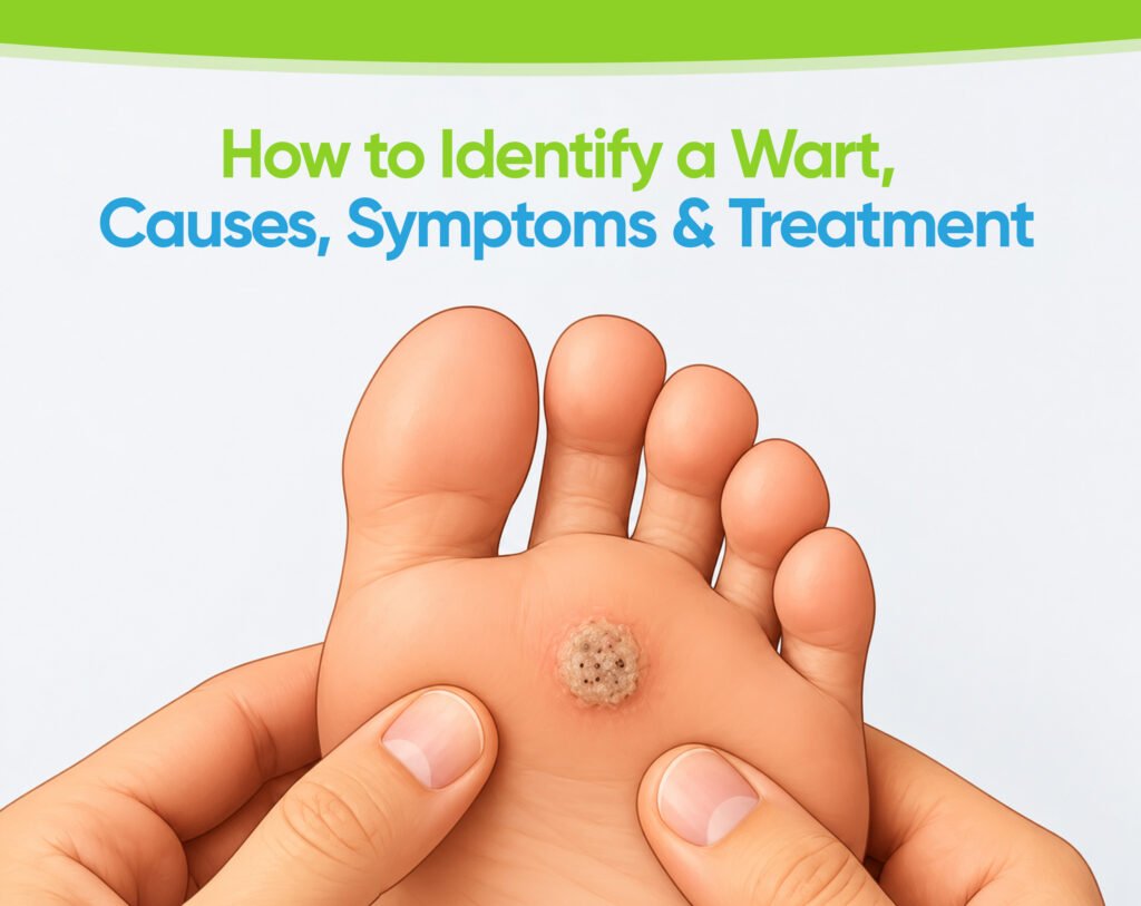
- “Wart Seeds” (Thrombosed Capillaries): Tiny black or dark red dots are often visible near the center of the growth. These are not true seeds; they are tiny, clotted blood vessels that supply oxygen and nutrients to the lesion.
Identifying the Primary Types of Warts
Dermatologists classify these benign epidermal growths based on their appearance and anatomical location.
1. Common Warts (Verruca Vulgaris)
- Location: Hands, fingers, around the nails, and knees.
- Appearance: Raised, rough bumps ranging from 1 to 10 millimeters in size. They often appear grayish-brown or flesh-colored with a hyperkeratotic surface.
2. Plantar Warts (Verruca Plantaris)
- Location: Soles of the feet, particularly pressure-bearing areas like the heels or balls of the feet.
- Appearance: Flattened rather than raised due to the pressure of walking. They grow inward into the thick stratum corneum, often causing pain similar to stepping on a small pebble. They typically exhibit multiple dark thrombosed capillaries in the center.
3. Flat Warts (Verruca Plana)
- Location: Face, neck, wrists, or legs (often spread by shaving).
- Appearance: Smooth, flat-topped, slightly raised lesions that are smaller (1 to 4 millimeters) and often occur in large numbers (20 to 100 at a time). They range in color from yellowish-brown to pink.
4. Periungual and Subungual Warts
- Location: Around (periungual) or underneath (subungual) fingernails and toenails.
- Appearance: Rough, uneven bumps that can alter nail growth or cause painful lifting of the nail plate.
Differential Diagnosis: How to Tell a Wart Apart from Other Skin Conditions
Misidentifying a lesion can lead to ineffective care. Use this comparison table to distinguish viral skin growths from lookalike conditions:
| Feature | Viral Wart | Callus or Corn | Skin Tag | Molluscum Contagiosum |
| Primary Cause | Viral (HPV) | Friction / Pressure | Friction / Genetics | Viral (Poxvirus) |
| Surface Texture | Rough, grainy, hyperkeratotic | Thick, smooth or hard center | Soft, smooth, pedunculated | Smooth, firm dome |
| Black Dots Present? | Yes (clotted capillaries) | No | No | No (central dimple instead) |
| Fingerprint Lines | Interrupted by lesion | Continuous over callus | N/A (hangs off stalk) | N/A |
| Tenderness | Painful when squeezed side-to-side | Painful when pressed directly | Painless unless twisted | Usually painless |
When to Seek Professional Dermatological Evaluation?
While many small viral lesions resolve spontaneously as the immune system recognizes the viral strain, professional assessment is recommended in several scenarios:
- Uncertain Diagnosis: If you are unsure whether the lesion is benign, a professional examination rules out malignant or atypical skin conditions.
- Pain or Location Issues: Lesions on the feet, under nails, or on the face require specialized management to avoid scarring or severe discomfort.
- Spreading or Persistent Lesions: Multiple surrounding lesions indicate active viral shedding that may benefit from clinical intervention.
- Compromised Immune System or Diabetes: Individuals with poor circulation or impaired immunity should avoid over-the-counter chemical treatments and seek clinical care.
For a detailed visual guide and clinical breakdown on differentiating benign skin growths, readers can explore the resource on how to identify a wart by Dermpro, which covers diagnostic criteria and treatment options in detail.
Practical Dos and Don’ts for Managing Suspected Warts
Recommended Practices:
- Keep the Area Covered: Placing a small adhesive bandage over the lesion reduces the risk of autoinoculation (spreading the virus to other areas of your body).
- Wash Hands Thoroughly: Clean your hands after touching or applying topicals to the lesion.
- Keep Feet Dry: Change socks daily and wear footwear in shared public showers or locker rooms to avoid reinfection.
Practices to Avoid:
- Do Not Pick or Scratch: Picking at the rough surface spreads viral particles to nearby skin micro-abrasions.
- Avoid Shaving Over Lesions: Razor blades spread the virus quickly, particularly on the face (flat warts) or legs.
- Never Use Nail Clippers for Removal: Attempting home surgical removal causes severe bleeding, secondary bacterial infection, and scarring.
Frequently Asked Questions
Why do warts have tiny black dots in them?
The tiny black dots are thrombosed (clotted) capillaries. As the viral lesion grows, it stimulates small blood vessels to supply it with nutrients. When these tiny vessels clot off near the surface, they appear as dark specks.
Can a callus turn into a wart?
No. Calluses are caused purely by mechanical friction and pressure, whereas warts are caused by an active viral infection (HPV). However, a viral lesion on the sole of the foot can accumulate calloused skin over top of it due to walking pressure.
How long do viral skin growths typically last if left untreated?
In healthy individuals, up to 60% of common viral lesions clear spontaneously within two years as the immune system develops targeted antibodies. However, treatment is often preferred to reduce discomfort and prevent spreading to others.
Are water warts the same as common warts?
No. “Water warts” refers to Molluscum contagiosum, which is caused by a poxvirus rather than HPV. Molluscum lesions appear as smooth, dome-shaped bumps with a characteristic central dimple (umbilication), rather than a rough, hyperkeratotic surface.
Summary
Accurately identifying a wart involves looking for key diagnostic signs: a rough cauliflower-like texture, interrupted skin ridges, distinct borders, and tiny black dots representing clotted capillaries. Differentiating viral lesions from calluses, corns, and skin tags ensures you choose appropriate, safe management strategies and avoid unnecessary skin damage.