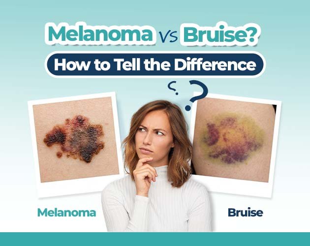
nail bruise vs melanoma
Discovering a dark spot, stripe, or discoloration beneath a fingernail or toenail is a common occurrence. In most cases, nail discoloration stems from minor trauma such as stubbing a toe or accidentally catching a finger in a door resulting in a subungual hematoma (a collection of blood under the nail).
However, dark nail pigmentation can occasionally signal a rare but serious form of skin cancer known as subungual melanoma. Because early detection significantly improves treatment outcomes, knowing how to distinguish a routine nail bruise from a suspicious lesion is vital.
This guide breaks down the clinical characteristics of both conditions, explaining how they behave over time and outlining when professional dermatological evaluation is necessary.
What Is Subungual Hematoma (Nail Bruise)?
A subungual hematoma occurs when trauma damages the blood vessels beneath the nail plate, causing blood to pool in the nail bed or matrix.
Common Causes
- Acute Trauma: A sudden impact, such as hitting a thumb with a hammer or dropping a heavy object on a foot.
- Micro-Trauma: Repetitive friction from tight footwear or activities like long-distance running and tennis.
Key Characteristics of a Bruise
- Color Evolution: Starts as reddish-purple or dark blue and gradually shifts to brown, black, or yellowish-green as the blood breaks down.
- Growth Behavior: As the nail plate grows outward, the discolored spot moves toward the free edge of the nail.
- Shape: Often appears as an irregular blotch or pool of color rather than a uniform vertical stripe.
- Sensation: Usually accompanied by localized tenderness or throbbing pain immediately after the injury, which diminishes over several days.
What Is Subungual Melanoma?
Subungual melanoma is a subtype of acral lentiginous melanoma, originating in the nail matrix ( the tissue beneath the cuticle where nail growth begins). Unlike typical skin cancers driven by ultraviolet (UV) radiation from sunlight, subungual melanoma is not linked to sun exposure.
It accounts for roughly 1% to 3% of cutaneous melanomas in light-skinned populations, but represents up to 20% to 30% of melanoma cases in individuals with darker skin phototypes. It occurs most frequently on the thumb, great toe, or index finger.
Key Characteristics of Subungual Melanoma
- Appearance: Typically presents as a vertical pigmented band (longitudinal melanonychia) running from the cuticle down to the tip of the nail.
- Color: Often dark brown or black, though amelanotic (non-pigmented) variants can appear pinkish or flesh-colored.
- Static Position: The pigment originates in the matrix, meaning the dark band persists and often widens over time rather than growing out with the nail.
- Lack of Pain: Early-stage lesions are almost always painless, which can delay diagnosis if the spot is mistaken for a bruise.
Comparison: Subungual Hematoma vs. Subungual Melanoma
Understanding the core visual and behavioral differences helps clarify whether a spot requires observation or immediate clinical evaluation.
| Clinical Feature | Subungual Hematoma (Nail Bruise) | Subungual Melanoma |
| History of Injury | Clear history of trauma or repetitive pressure | Usually no remembered injury |
| Growth Pattern | Moves outward as the nail grows | Remains fixed at the matrix base; may widen |
| Pigment Shape | Pool, blotch, or irregular spot | Vertical stripe running from cuticle to tip |
| Hutchinson’s Sign | Absent (pigment stays within nail plate) | Often present (pigment spills onto cuticle skin) |
| Nail Changes | Nail remains intact (unless severe trauma) | Nail plate may split, crack, or lift (onycholysis) |
| Pain Level | Painful initially, then fades | Typically painless in early stages |
Diagnostic Clues: The ABCDEF Rule for Nail Melanoma
Dermatologists frequently use the specialized “ABCDEF” clinical framework to assess pigmented nail streaks for potential malignancy:
- A – Age: Most commonly diagnosed in adults between 50 and 70 years old.
- B – Band Color and Width: A dark brown or black band that is wider than 3 millimeters or displays irregular color shading.
- C – Change: Changes in the width, shape, or color of the nail streak over time.
- D – Digit Involved: Most frequently affects the thumb, big toe, or index finger.
- E – Extension (Hutchinson’s Sign): Pigment spreading beyond the nail plate onto the surrounding cuticle or proximal nail fold.
- F – Family History: Personal or family history of dysplastic nevi or cutaneous melanoma.
Visual Indicator to Watch For:
[Cuticle Skin] ──(Pigment Spills Over)──> [Hutchinson’s Sign = Suspicious]
[Nail Plate] ──(Vertical Dark Line)──> [Requires Evaluation]
When Should You See a Doctor?
While the vast majority of dark nail spots are benign bruises, certain signs warrant a professional skin check. A dermatologist uses dermoscopy, a specialized magnifying tool with polarized light to non-invasively examine the pigment patterns in the nail matrix.
Schedule an Evaluation If You Notice:
- A dark line under your nail that appeared without any trauma.
- A spot or streak that widens, darkens, or changes shape over weeks.
- Pigment that extends onto the skin surrounding the nail bed.
- A nail plate that begins to split, crack, or separate from the nail bed.
- A bruise that does not grow outward or change position after 6 to 8 weeks.
To understand how clinical evaluations, dermoscopic exams, and biopsies are conducted when assessing suspicious nail changes, reviewing the comprehensive guide on melanoma from nail bruises at Sayyal Health provides helpful context on clinical diagnostic pathways.
Frequently Asked Questions
How long does a nail bruise take to grow out fully?
Because fingernails grow at an average rate of 3 millimeters per month (and toenails grow even slower), a subungual hematoma can take 6 to 9 months to completely grow out on a finger, and up to 12 to 18 months on a toe.
Can a nail bruise turn into melanoma?
No. A subungual hematoma is simply trapped blood under the nail plate resulting from vascular damage. Trauma does not cause cellular DNA mutations that lead to malignant melanoma. However, an early melanoma lesion can easily be misdiagnosed as an accidental injury.
What does a dermatologist do if they suspect melanoma under a nail?
If dermoscopy reveals suspicious pigment patterns, the dermatologist will perform a nail matrix biopsy. This involves numbing the digit and taking a small tissue sample from the matrix for histopathological analysis to check for atypical melanocytes.
Can fungal infections cause dark spots under the nail?
Yes. Certain fungal organisms (like Trichophyton rubrum) or non-fungal molds can produce dark pigments (melanonychia) that resemble bruises or melanomas. A physician can perform a fungal culture or clipping to rule out infection.
Summary
Differentiating a common nail bruise from a rare subungual lesion comes down to tracking growth behavior, color stability, and pattern. A bruise will move outward as the nail grows and fade over time, whereas suspicious pigmentation persists at the cuticle base and may widen or spill onto surrounding skin. If you observe an unexplained dark streak that fails to grow out, consulting a healthcare professional ensures peace of mind and timely care.