Australia has one of the highest rates of skin cancer and related sun-damage conditions in the world. Living under the harsh southern sun means that our skin bears a cumulative burden of ultraviolet (UV) radiation. Over time, this chronic exposure can manifest as rough, dry, or scaly patches known medically as Actinic Keratosis (and often referred to as solar keratosis).
While these spots might initially seem like minor cosmetic annoyances or stubborn patches of dry skin, they are actually precancerous lesions. If left unmanaged, a percentage of these patches can progress into a type of skin cancer known as Squamous Cell Carcinoma (SCC). Understanding how to spot the early warning signs, recognizing the underlying causes, and exploring modern clinical treatments is crucial for safeguarding your long-term health.
This comprehensive guide, backed by Australian dermatological contexts and clinical insights from dermpro.com.au, breaks down everything you need to know about managing this highly prevalent condition.
What is Actinic Keratosis?
Actinic Keratosis is a common, precancerous skin growth caused by long-term exposure to ultraviolet (UV) radiation. It typically develops on chronically sun-exposed areas of the body, such as the face, lips, ears, bald scalp, neck, and the backs of the hands or forearms.
The condition occurs when the DNA inside our epidermal skin cells, specifically the cells called keratinocytes, becomes permanently altered by UV rays. This cellular damage causes the cells to grow abnormally, resulting in rough, scaly lesions.
Why It’s Considered a Precursor to Skin Cancer
Statistically, around 40% to 50% of fair-skinned Australian adults over the age of 40 will develop at least one Actinic Keratosis lesion. While an individual spot has a low annual rate of transformation, clinical data shows that 60% to 65% of all Squamous Cell Carcinomas arise directly from a pre-existing Actinic Keratosis patch. Because it is impossible to predict which specific spots will advance into an invasive malignancy, medical professionals strongly recommend tracking and treating them early.
Symptoms: How to Identify Solar Keratosis?
Identifying an Actinic Keratosis spot can sometimes be tricky because they often present more as a specific physical texture rather than a highly visible mark. Many people actually feel them before they see them.
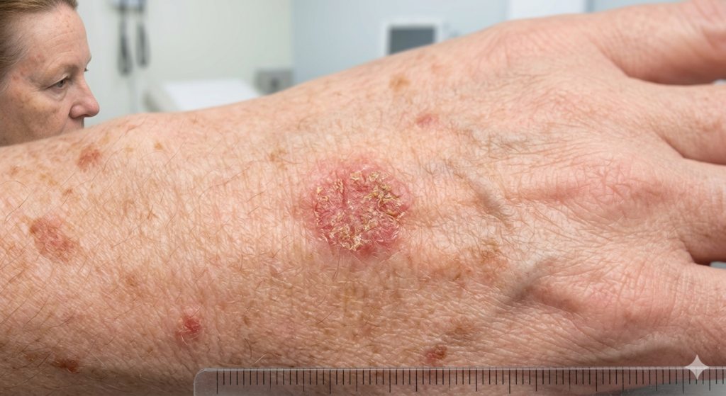
What Does Actinic Keratosis Feel and Look Like?
- The Sandpaper Texture: The most definitive clinical sign is a rough, gritty surface. Rubbing your finger over the spot feels remarkably like running it across fine sandpaper.
- Visual Variations: They can appear as flat or slightly raised patches. While many are pink, red, or flesh-colored, some can look light grey, yellowish, or brown.
- Persistent Scales: They frequently produce a dry, crusty, or scaly layer. If you pick the scale off, it will almost always grow back in the exact same spot within a few weeks.
- Localized Sensation: The area may occasionally itch, burn, sting, or feel tender and tender to the touch, especially when exposed to direct sunlight or rubbed by clothing.
Clinical Grades of Severity
Dermatologists categorize these lesions into three distinct visual and tactile grades:
- Grade 1 (Mild): Flat, faint pink or grey marks that are barely visible but easily felt as a slight, gritty texture.
- Grade 2 (Moderate): Moderately thick, rough, hyperkeratotic patches that are distinctly visible and easily detected during a skin examination.
- Grade 3 (Severe): Thick, heavily scaling, or hyperkeratotic growths. These can sometimes produce an excess of hard keratin, forming a pronounced, cone-like protrusion known as a cutaneous horn.
Primary Causes and Risk Factors
The fundamental, driving cause of Actinic Keratosis is cumulative exposure to ultraviolet (UV) radiation. This includes both natural sunlight and artificial UV light from indoor tanning beds.
When UV rays penetrate the outer layer of the skin, they break down cellular DNA bonds. While the body has built-in mechanisms to repair this damage, decades of intense sun exposure overwhelm these defense pathways. The damaged cells begin to replicate out of control, surfacing as rough patches years or even decades after the initial sun exposure took place.
Who is Most at Risk?
While anyone can develop solar keratoses, certain demographic and lifestyle factors significantly elevate your risk profile:
- Skin Phenotype: Individuals with fair skin (specifically Fitzpatrick Skin Types I and II), red or blonde hair, light-colored eyes, and a tendency to freckle or burn easily.
- Age and Cumulative Exposure: Because the UV damage is cumulative, prevalence climbs dramatically with age. It affects roughly 10% of people in their 20s, but rises to over 90% in individuals over 80.
- Geographic Location: Living in regions with high UV indexes year-round, such as Queensland or northern territories of Australia, drastically accelerates the timeline of skin damage.
- Immune Status: People with compromised immune systems such as organ transplant recipients on immunosuppressant medications have a substantially higher rate of developing multiple, aggressive lesions.
- Occupational and Recreational Roles: Outdoor workers (tradies, farmers, landscapers) and people who frequently participate in open-air sports (golfing, surfing, cricket) experience a baseline of chronic UV exposure that drives up risk.
1. Lesion-Directed Therapies
- Cryotherapy: This is the most widely utilized treatment in Australian general practices. A clinician applies liquid nitrogen to freeze the targeted spot. The extreme cold destroys the abnormal keratinocytes, causing the area to blister, scab, and flake away as healthy new skin regenerates underneath.
- Curettage and Cautery: Used primarily for thick, advanced Grade 3 spots. The doctor numbs the skin, scrapes away the abnormal tissue using a sharp tool called a curette, and applies heat (cautery) to stop bleeding. This allows the doctor to send a tissue sample away for a biopsy to completely rule out invasive skin cancer.
2. Field Cancerisation Treatments
Often, the apparently healthy skin surrounding a visible lesion has undergone the exact same degree of underlying UV damage. This is known as “field cancerisation.” If left untreated, new spots will continually pop up in that zone.
- Prescription Topical Creams: Medicated ointments are applied to an entire area over a set period. Common options include 5-Fluorouracil (5-FU), a localized chemotherapy cream that kills rapidly dividing cells, and Imiquimod, an immunotherapy cream that prompts your body’s own immune system to target and destroy the precancerous tissue. A newer, short-course option is Tirbanibulin, which requires only a 5-day application cycle.
- Photodynamic Therapy (PDT): A specialized photosensitizing cream is applied to the skin and left to absorb for a few hours, where it selectively builds up in the abnormal cells. The area is then exposed to a specific wavelength of light (or natural daylight in “Daylight PDT”), activating the cream and destroying the damaged cells while preserving the underlying healthy structure.
Prevention and Long-Term Management
The most effective way to prevent new Actinic Keratosis lesions from forming and to reduce the risk of recurring spots after a treatment cycle is strict, daily UV protection.
- Broad-Spectrum Sunscreen: Apply a therapeutic, broad-spectrum sunscreen with an SPF rating of 50+ every morning to all exposed skin. Reapply every two hours if you are outdoors or swimming. Consistent sunscreen use has been clinically shown to allow some early-stage, flat lesions to regress naturally.
- Physical Barriers: Wear sun-protective clothing, including tightly-woven fabrics, long sleeves, a wide-brimmed hat that shields the face, ears, and neck, and UV-blocking sunglasses.
- Peak Hour Avoidance: Stay in the shade during peak UV hours, typically between 10:00 am and 4:00 pm, when the solar radiation index is at its highest.
- Nicotinamide (Vitamin B3): Clinical research in Australia suggests that taking a specific dose of Nicotinamide (500 mg twice daily) can help support cellular DNA repair mechanisms, effectively reducing the formation of new solar keratoses and non-melanoma skin cancers in high-risk individuals. Always consult your GP before starting new supplement regimens.
Frequently Asked Questions
Is Actinic Keratosis considered cancer?
No, Actinic Keratosis is not cancer. It is classified as a precancerous or premalignant skin lesion. This means the cellular changes are currently confined to the very top layer of the skin (the epidermis). However, if left untreated over a long period, a small percentage of these lesions can break through the basement membrane and progress into an invasive form of skin cancer called Squamous Cell Carcinoma.
Can an Actinic Keratosis spot go away on its own?
Yes, some mild, early-stage Actinic Keratosis lesions can resolve on their own, particularly if you implement rigorous, daily sun protection. When you consistently protect the skin from UV rays, it gives your body’s natural cellular repair mechanisms an opportunity to clear the damaged cells. However, many spots that appear to disappear are simply dormant and will scale up again when exposed to UV light, which is why clinical monitoring is recommended.
How often should I get my skin checked if I have these spots?
If you have been diagnosed with Actinic Keratosis, it indicates that your skin has reached a threshold of cumulative sun damage. As a result, you are at a higher baseline risk for developing other forms of skin cancer. You should perform self-examinations at home monthly, and visit a dermatologist or dedicated skin cancer clinic at least once a year for a comprehensive, professional full-body skin check.
What happens if you pick off the crust of a solar keratosis?
Picking the scaly crust off an Actinic Keratosis lesion will not cure it. The rough surface is merely an outward symptom of abnormal cell growth occurring deeper within the epidermis. If you force the scab or scale off, the area will typically bleed, feel tender, and eventually grow the exact same rough, sandpaper-like texture back within a couple of weeks once the skin surface closes.
Do topical treatments for Actinic Keratosis leave scars?
Field treatments like 5-Fluorouracil, Imiquimod creams, or Photodynamic Therapy (PDT) rarely leave permanent scars because they target abnormal cells while leaving the healthy skin infrastructure intact. However, during the treatment process, you will experience a significant, temporary local inflammatory reaction involving redness, peeling, crusting, and swelling. This is a normal sign that the therapy is working. Once healed, the skin is usually smoother and healthier.
Conclusion
Actinic Keratosis is a clear, physical warning sign from your body that your skin has sustained significant, cumulative UV damage. While these rough, sandpaper-like patches are not yet fully developed skin cancer, their strong evolutionary link to Squamous Cell Carcinoma means they should never be ignored.