When a dark spot appears under a fingernail or toenail, the immediate reaction is often a mix of confusion and anxiety. Most people assume they simply slammed their finger in a door or dropped something heavy on their toe, resulting in a common nail bruise.
However, an identical dark line or spot can also be the initial presentation of melanoma of the nail unit specifically, a rare but serious variant known as subungual melanoma.
Because a malignant lesion can masquerade perfectly as an everyday injury, understanding the clinical difference is vital. Failing to recognize the distinct signs of cancer on nails can lead to delayed diagnoses, while confusing a simple bruise for a malignancy causes unnecessary panic.
What is Subungual Melanoma?
Subungual melanoma is a form of skin cancer that originates within the nail matrix the tissue directly under the nail piece where new nail growth forms. Unlike traditional cutaneous melanomas, which are heavily driven by ultraviolet (UV) radiation from sun exposure, subungual melanoma is largely unrelated to sunlight. Instead, it falls under the category of acral lentiginous melanoma, a subtype that develops on non-sun-exposed areas like the palms of the hands, soles of the feet, and nail beds.
While melanoma of the nail unit accounts for only about 1.9% to 3% of all cutaneous melanoma cases in fair-skinned populations, it represents a disproportionately higher percentage of melanoma diagnoses in individuals with darker skin tones, including those of African, Asian, and Hispanic descent. It most frequently affects the thumb and the great toe, though it can develop on any digit.
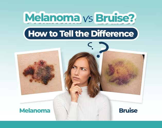
Nail Bruise vs Melanoma: Visual Differences
Distinguishing between a standard trauma-induced hematoma (blood pool) and a malignant growth requires careful evaluation of how the pigment behaves over time.
The Nature of a Nail Bruise (Subungual Hematoma)
A subungual hematoma occurs when trauma damages the blood vessels beneath the nail plate, causing blood to pool between the nail bed and the plate.
- Appearance: It typically presents as a discrete, sharp-edged discoloration that varies from bright red to purple, maroon, or jet black, depending on the age of the trapped blood.
- Growth Pattern: Because the blood is physically trapped on top of the nail bed, the spot will move upward naturally as your nail grows out. Over several months, a clear gap of normal, unpigmented nail will emerge at the base (cuticle) until the bruise eventually reaches the tip and is clipped away.
The Nature of Subungual Melanoma
When malignant melanocytes (pigment-producing cells) in the nail matrix begin to proliferate abnormally, they continuously deposit melanin into the newly forming nail plate.
- Appearance: This creates a vertical band of pigment known as longitudinal melanonychia. Rather than a stagnant blob of blood, it looks like a continuous, deliberate stripe running from the cuticle line straight to the free edge of the nail.
- Growth Pattern: Because the source of the pigment is actively growing at the root, the stripe does not clear out or move away from the cuticle. Even as the nail grows, the dark band remains fixed, often widening or changing colors over time.
When Does a Bruise Warrant Medical Evaluation?
It is surprisingly common for individuals diagnosed with subungual melanoma to recall an isolated incident of trauma such as dropping an object on their foot or stubbing a toe prior to the spot appearing. This correlation frequently leads to a dangerous assumption that the spot is entirely benign, delaying necessary evaluation.
Trauma can cause a genuine bruise, but a physical injury can also draw a patient’s attention to a pre-existing, unnoticed malignant line. Furthermore, chronic mechanical stress or severe acute trauma have been discussed as potential co-factors that disrupt tissue microenvironments.
The 8-Week Standard Rule: If you notice a dark spot on your nail that you suspect is a bruise, monitor it closely. If the discoloration shows absolutely no outward movement away from the cuticle line after 6 to 8 weeks, or if it continues to widen and darken, you must seek a professional dermatological assessment.
Professional Diagnosis and Treatment Pathways
When you consult a medical professional regarding a suspicious nail line, the diagnostic process follows a strict, safe hierarchy to prevent unnecessary surgery while ensuring malicious cells are caught early.
1. Advanced Dermoscopy (Onychoscopy)
A dermatologist will initially examine the nail using a high-powered, polarized magnifying tool called a dermoscope. Under dermoscopy, a standard nail bruise reveals characteristic reddish-black homogeneous puddles, round structures called globules, or peripheral fading where the blood is thinning out. Conversely, subungual melanoma displays a chaotic arrangement of parallel longitudinal lines that vary in thickness, spacing, and coloration.
2. Full-Thickness Nail Biopsy
If dermoscopy reveals irregular vascular patterns or structural chaos, a biopsy remains the absolute gold standard for diagnosis. Performed under a localized digital nerve block to fully numb the area, a surgeon will gently remove a tiny portion of the nail plate to harvest a small tissue sample from the underlying nail matrix or bed.
3. Surgical Intervention
If histopathological analysis confirms the presence of subungual melanoma, treatment relies primarily on surgical excision.
- Early-Stage/In-Situ: For early-stage melanomas confined strictly to the upper layers of the tissue, modern practice prioritizes complete functional preservation by performing a wide local excision of the entire nail apparatus followed by a skin graft, avoiding amputation entirely.
- Invasive/Advanced: For deep, invasive lesions that have penetrated down toward the underlying bone, a partial or complete digit amputation may be required to achieve clear, cancer-free margins and preserve long-term health.
Frequently Asked Questions
Can a nail bruise look exactly like melanoma?
Yes. An atypical subungual hematoma especially one caused by repetitive micro-trauma from tight footwear can present as a dark, linear streak that closely mimics the signs of cancer on nails. Professional dermoscopy is usually required to visualize the microscopic structural differences between pooled blood and melanin clusters.
How long does it take for a nail bruise to grow out completely?
Fingernails grow at an average rate of 3 millimetres per month, meaning a bruise can take anywhere from 4 to 6 months to grow out completely. Toenails grow much slower, averaging about 1 millimetre per month, meaning a deep toe bruise can take a full 12 to 18 months to clear.
Does subungual melanoma cause pain or nail splitting?
In its early stages, subungual melanoma is completely painless and presents strictly as a visual color change. However, as the tumor expands within the delicate nail matrix, it disrupts normal nail production, which can cause the nail plate to split vertically, crack, lift from the nail bed, or develop chronic ulceration and discharge